
Figure 1.
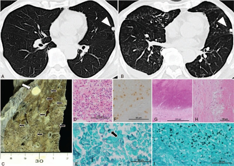
Chest computed tomography images revealing a solitary pulmonary nodule in the lingular segment (arrowhead) 5 years prior to the currently discussed admission (A), randomly distributed diffuse small nodular shadows at the time of admission (B). Macroscopic findings from the lingular segment of the lung revealed a calcified nodule (white arrow) and small necrotizing lesions (black arrow) (C). Microscopic analysis of the diffuse small nodular lesions revealed granulomatous areas with necrosis (D) and yeast-like fungi detected using Grocott stain (black arrow) (E) and immunohistochemistry with a Histoplasma-specific antibody (Meridian Bioscience) (F). The calcified nodule underlying the visceral pleura was surrounded and coated with fibrous material (G) and exhibited necrosis (H) and many yeast-like fungi (I).