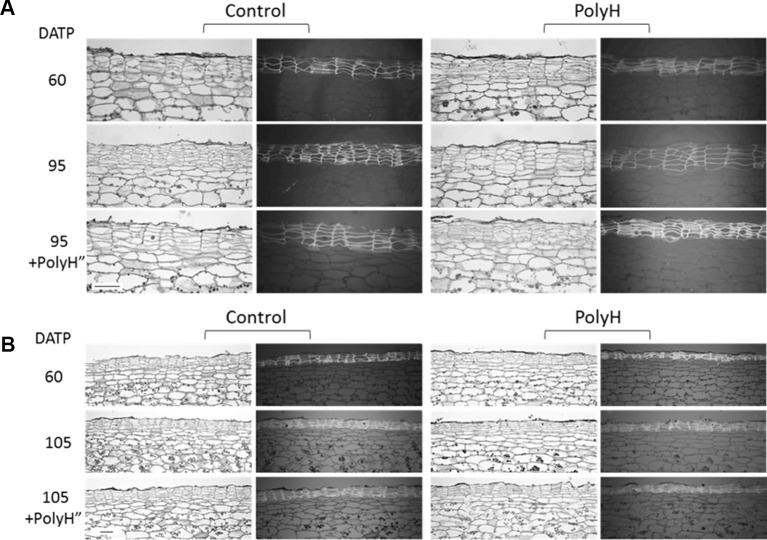
Figure 9.
Anatomical study of tuber skin of potato cvs. Rosanna (A) and Vivaldi (B) (experiment 3). Sample labeling and treatments are as described in Figures 5 and 6 . Sections were taken from three independent tubers for each treatment and developmental stage, and one representative section is shown. Each section is represented by two frames: the left shows tissue morphology under a light microscope, the right shows the autofluorescence of skin cells on a dark gray background as seen by UV microscope. Note that the morphology of the potato skin is characterized by cell column arrangements. Bar = 100 µm.

