Figure 3.
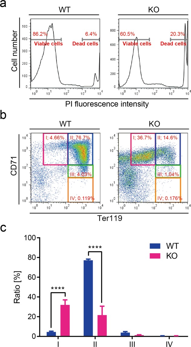
Flow cytometric analysis of liver cells from wild-type and Shmt2-knockout E13.5 embryos. (a) Dead cells from wild-type and Shmt2-knockout foetal livers were stained with PI and separated from viable cells using flow cytometric analysis. The same number (3 × 10⁴) of viable cells were then analysed using CD71/Ter119 flow cytometric assay. Representative results (b) and quantitative evaluation (c) of flow cytometric analysis of foetal liver cells. In the E13.5 embryonic stage, foetal livers function as erythropoietic sites, with most cells in the foetal liver representing progenitors of erythrocytes. In wild-type embryos (embryos without deficient Shmt2), most progenitors are at stage II (a CD71High/Ter119High subset mostly consisting of erythroblasts). In Shmt2-knockout embryos, >30% of cells are at the undifferentiated stage I (a CD71High/Ter119Middle subset mostly consisting of pro-erythroblasts), indicating the arrest of erythroblast differentiation. Cells further along in differentiation [i.e., stage III (a CD71Middle/Ter119High subset mostly consisting of reticulocytes) and stage IV (a CD71Low/Ter119High subset mostly consisting of matured erythrocytes)], are rare, even in wild-type embryos. WT, wild-type foetal livers (solid blue bars); KO, Shmt2-knockout foetal livers (solid red bars). Data represent the mean ± S.D. ****P < 0.0001, 2-way ANOVA (WT, n = 3; KO, n = 4).

