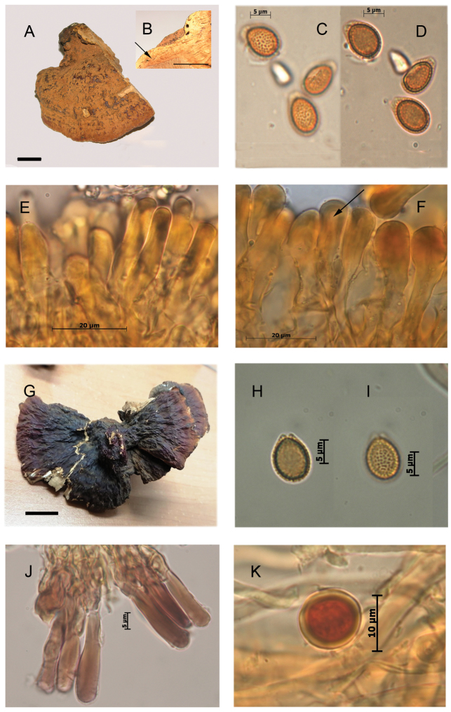
Figure 7.
Morphological features and microscopic structures of Ganoderma sessiliformeA–F NY 98713 (holotype) A pilear surface B context not fully homogeneous with discrete bodies of the resin-like deposits (arrow) C–D basidiospores with free to subfree pillars E–F cuticular cells in KOH E cylindrical F clavate, with narrow lumen (arrow) G–K Guzmán 2078, ENCB G pilear surface H–I basidiospores with free to subfree pillars J cuticular cells in Melzer reagent K smooth-walled chlamydospore from context in Melzer reagent.

