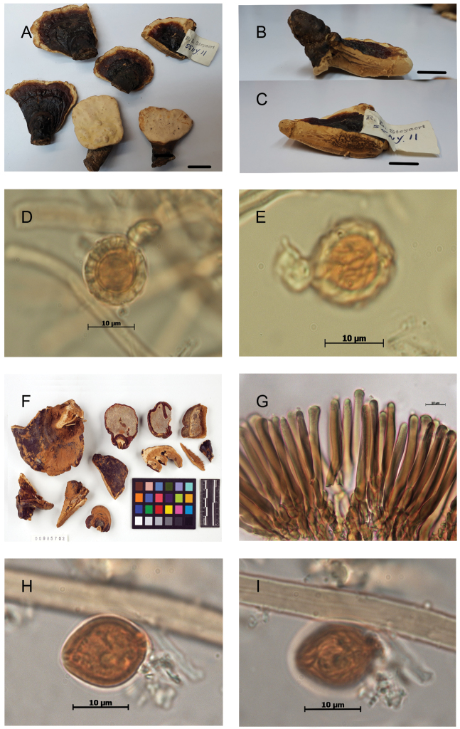
Figure 8.
Morphological features and microscopic structures of type specimens of GanodermaA–EGanoderma stipitatum (NY 985678, holotype) A basidiomata B–C context B brown stripes of resinous deposits C numerous bodies of the resin-like deposits D–E chlamydospore, ornamented with partially anastomosed ridges, from context F–IGanoderma perzonatum (NY 985702, holotype), F basidiomata, copyright: NY Botanical Garden G cuticular cells, cylindrical, with incrustations H–I chlamydospore with fine longitudinal ridges, from context. Scale bar: 1 cm (A–C).

