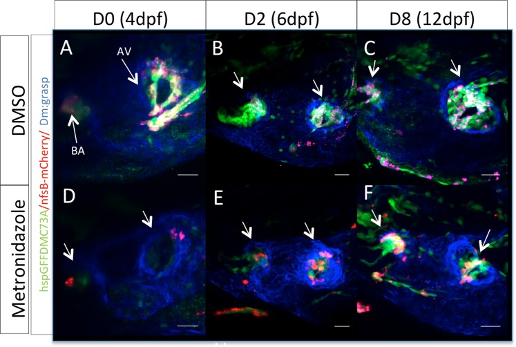
Figure 2.
Zebrafish embryonic valves can regenerate. (A–C) Confocal z-stacks of untreated Tg(hspGFFDMC73A/UAS-E1b:NfsB-mCherry) larvae at 4 dpf, 6 dpf and 12 dpf respectively. (D–F) Confocal z-stacks of MTZ treated Tg(hspGFFDMC73A/UAS-E1b:NfsB-mCherry) and larvae at 4 dpf, 6 dpf and 12 dpf respectively. Arrows show the ablation of mCherry+ cells at the AV canal and OFT at 4 dpf. From 6 dpf more mCherry+ cells could be observed at the AV canal. By 12 dpf the regeneration process could be imaged, as mCherry+, AV differentiated cells were present at both valves. Scale bars: 20 microns. Each experiment was carried out at least 3 independent times with n = 10 embryos per experimental group.

