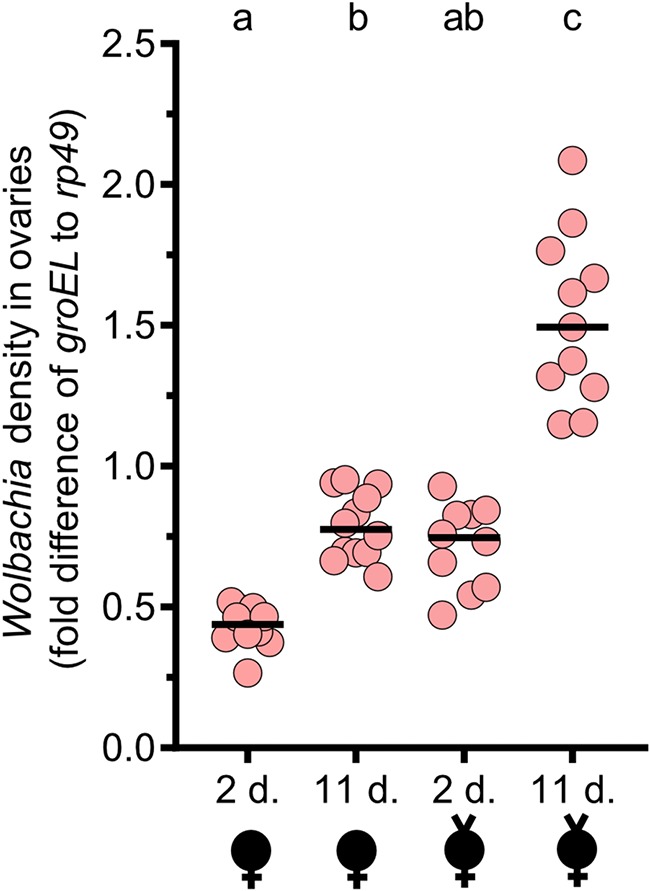
FIG 3.

Wolbachia densities increase with female age in ovaries and decrease after mating. Wolbachia density assays were conducted with pools of 4 ovaries from virgin females (indicated by a “v” above a sex symbol) and nonvirgin females aged 2 or 11 days (d.). Wolbachia infections are represented by filled sex symbols, and the age of the sample is shown immediately below the x axis. Virgin and nonvirgin females were siblings. The nonvirgin females produced the embryos whose results are shown in Fig. 2B. Nonvirgin females were allowed to mate and lay for 48 h before ovary dissections. Nonvirgin and virgin females were incubated for that same period of time and dissected in parallel. Each dot represents the average of duplicate values. Horizontal bars indicate medians, and the letters above the bars indicate significant differences based on α = 0.05 calculated by a Kruskal-Wallis test followed by a Dunn’s multiple-comparison test performed between all groups. All statistical values are presented in Table S1. Fold differences in Wolbachia density (groEL) relative to D. melanogaster reference gene rp49 were determined with 2−ΔΔCT.
