
Figure 5.
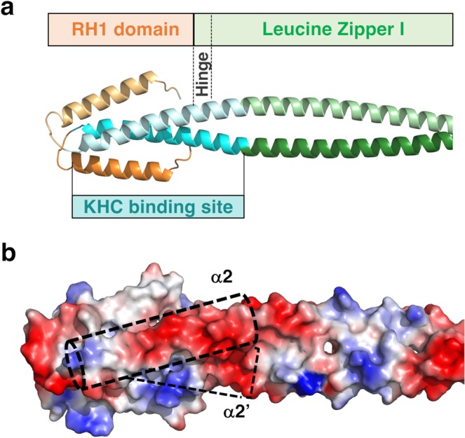
KHC-binding site overlaps with the RH1 domain of JIP3/4. (a) Homology model of JIP3-RH1-LZI tandem is shown in cartoon with the same color code as in Fig. 1 and the KHC-binding region (aa. 50–80) indicated in cyan. Only the first six heptad repeats of LZI are shown for clarity. (b) A surface representation of the JIP3-RH1-LZI model is shown in the same view as in (a). Acidic and basic residues are colored in red and blue, respectively; other residues are colored in white. The position of KHC-binding sites (aa 50–80) are indicated in dashed cyan boxes.