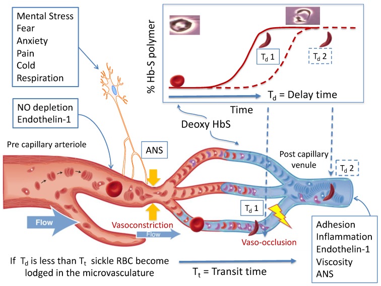
Figure 1.
The basic pathophysiological model of sickle vaso-occlusion suggests that microvascular occlusion will occur if the delay time from deoxygenation of HbS to polymerization (Td 1) is shorter than the microvascular transit time (Tt), as depicted in the lower two vessels. If delay time is longer (Td 2), or if blood is flowing faster, the red cell transition to a rigid shape takes place in a larger diameter post capillary vessel and occlusion is less likely to occur, as depicted in the top micro-vessel. Processes like nitric oxide (NO) depletion and endothelin-1 levels in the precapillary arterioles, and adhesion, inflammation, and viscosity in the post-capillary venule establish a steady state microvascular flow “tone”. Autonomic nervous system (ANS)-mediated vasoconstriction can decrease blood flow within seconds, increasing transit time and the likelihood of entrapment of rigid RBC. Image reprinted with permission from [19].

