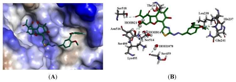
Figure 8.
The docked configuration of (+)-17b in the binding site of Tdp1 as predicted using the ChemScore scoring function. (A) The protein surface is rendered. The ligand occupies the binding pocket. Blue depicts a hydrophilic region on the surface; brown depicts hydrophobic region and whites shows neutral areas. (B) Hydrogen bonds are shown as green lines between Thr261, Asn516. The fluoride moiety interacts with Gln241 and His237. The water molecules also form hydrogen bonds with Ser459, Ser514, and Lys495.

