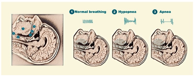
Figure 5.
Image of the upper airway seen by magnetic resonance and its collapsibility in patients with OSA and anatomical description of the upper airway (left); representation of (1) normal breathing: 1: the pharynx; 2: the larynx; 3: the genioglossus muscle; 4: the epiglottis; 5: the hard palate; and 6: the soft palate; (2) partial upper airway obstruction; and (3) complete obstruction of the upper airway.

