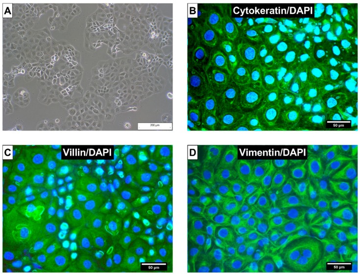
Figure 2.
(A) Morphology of calf small intestinal epithelial cells B (CIEB) visualized with inverse light microscopy (passage 10, 100 × magnification). Immunostaining of CIEB in chamber slides with (B) cytokeratin as an epithelial cell marker, (C) villin as marker for intestinal cells, and (D) vimentin as mesenchymal marker. 4′,6-Diamidin-2-phenylindol (DAPI) was used as cell nuclei counterstain (400× magnification).

