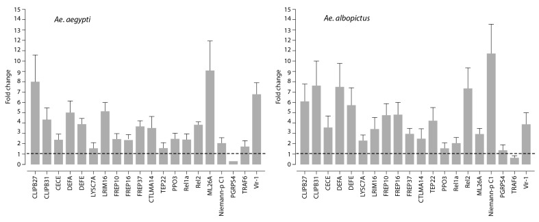
Figure 4.
Quantification of immune gene expression. Fold change in putative immune gene expression in the midgut following the feeding of Ae albopictus and Aedes aegypti mosquitoes with MAYV-containing blood, as compared to non-infectious blood meal as a control. The gene expression profile was determined by real-time PCR at 3 days post-blood-feeding. The gene expression of mosquitoes that were fed with non-infected blood meal as a control was represented as 1 (baseline). Experiments were performed three times, and error bars represent standard error of the mean.

