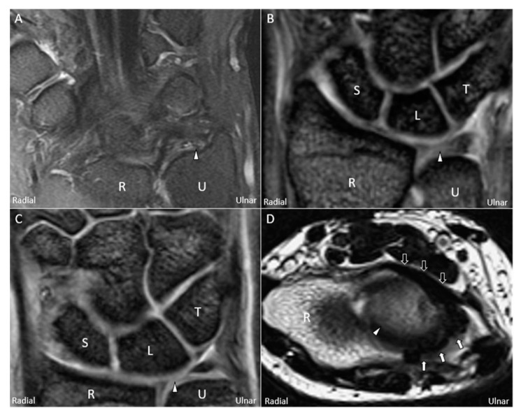
Figure 2.
Magnetic resonance imaging (coronal T2-weighted view) showing (A) avulsion of the articular disc (white arrowhead) from the ulnar fovea, (B) central tear of the disc, and (C) radial avulsion of the articular disc. (D) Magnetic resonance imaging (axial T2-weighted view) showing a swollen palmar radioulnar ligament (black arrow) and a torn dorsal radioulnar ligament (white arrow) surrounding the articular disc. U: ulna; R: radius; L: lunate; T: triquetrum; S: scaphoid.

