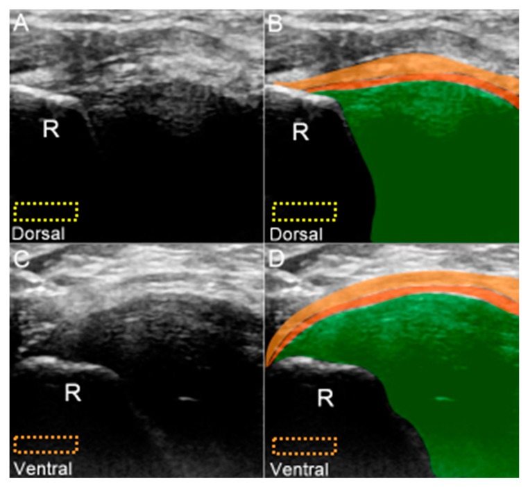
Figure 7.
(A) US and (B) superposed US images of the dorsal radioulnar ligament. (C) US and (D) superposed US images of the ventral radioulnar ligament. The different color dashed rectangles are in accordance with the transducer positions in Figure 3. US: ultrasound; R: radius. Superficial limb of the radioulnar ligament (light brown shade), deep limb of the radioulnar ligament (dark brown shade), and articular disc (green shade).

