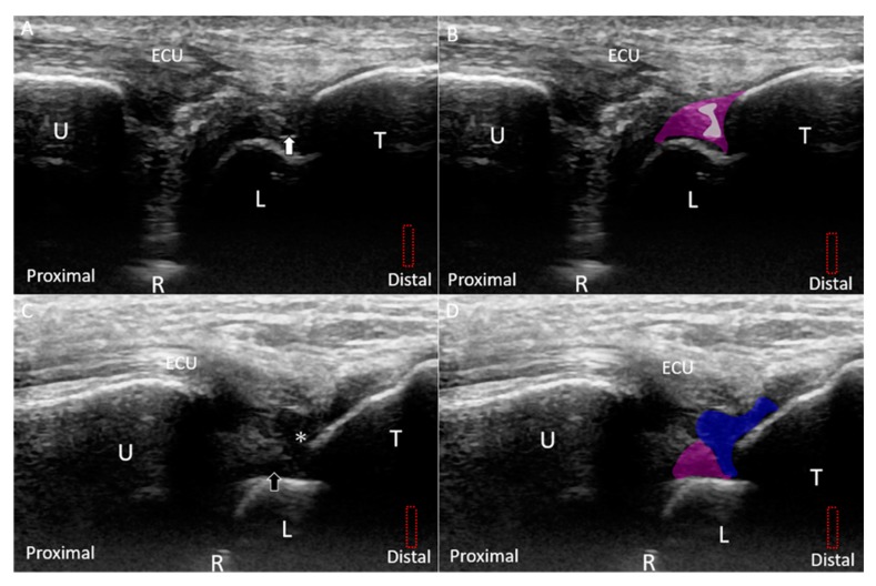
Figure 14.
(A) US and (B) superposed US images show a small tear (white arrow and grey shade) in the lunotriquetral ligament (purple shade) under the long-axis approach. (C) US and (D) superposed US images of the retracted stump (white asterisk and blue shade) of the torn lunotriquetral ligament and fluid accumulation (black arrow and purple shade). The different color dashed rectangles are in accordance with the transducer positions in Figure 3. US: ultrasound; U: ulna; R: radius; L: lunate; T: triquetrum; ECU, extensor carpi ulnaris.

