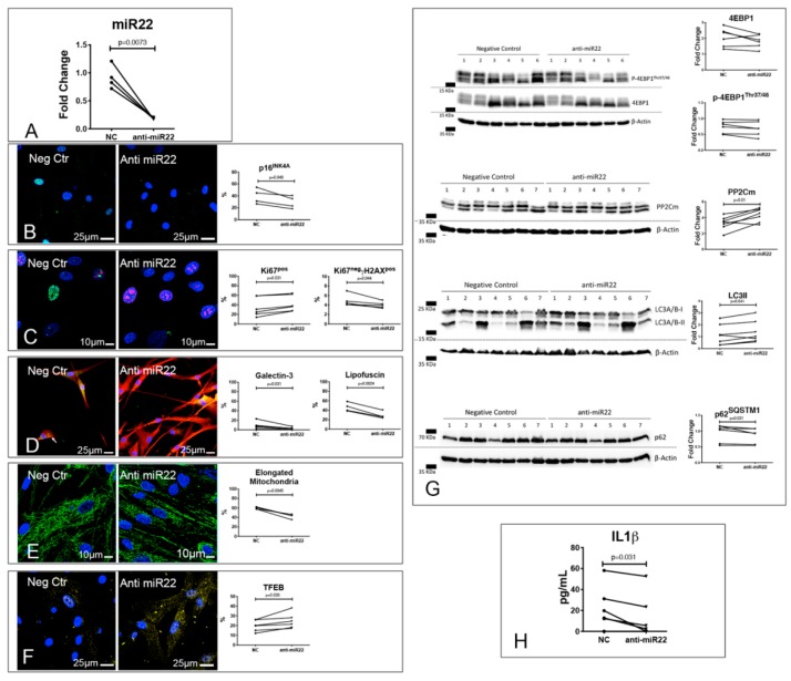
Figure 6.
Inhibition of miR-22 reverses EDCM-CPc senescence. Results of RT-PCR analysis of EDCM-CPc treated with either anti-miR22 or a negative control (NC; (A)). (B–F) Reversal of EDCM-CPc senescence and ALP dysfunction by miR-22 inhibition. Confocal images of EDCM-CPc treated with either anti-miR22 or NC showing positivity to p16INK4A (green, (B)), γH2A.X (green) and Ki67 (red, (C)), lipofuscins (green) and Galectin 3 (red, D), mitochondria (green, (E)), and TFEB (yellow, (F)). Nuclei are labeled by DAPI in blue. (G) Effects of miR-22 inhibition on mTOR targets, PP2Cm, and autophagy. Representative WB of whole protein extracts of 6–7 EDCM-CPc treated with either anti-miR22 or NC (G). Blotted proteins were incubated with antibodies against 4EBP1, p-4EBP1Thr37/46, PP2Cm, LC3A/B or p62SQSTM1. Results of the quantitative analyses are reported in the right panels. (H) Effects of miR-22 inhibition on IL1β secretion. ELISA assay was used for the detection of IL1β released in the culture supernatant of 7 EDCM-CPc treated with either anti-miR22 or NC (H). Dot plots indicate the results of quantitative analyses; paired t tests (p16INK4A, Ki67, lipofuscin, mitochondria, TFEB, 4EBP1, p-4EBP1Thr37/46, PP2Cm, and LC3A/B) or Wilcoxon tests (Ki67−γH2A.X+, Galectin 3, p62SQSTM1, and IL1β) were employed, as appropriate.

