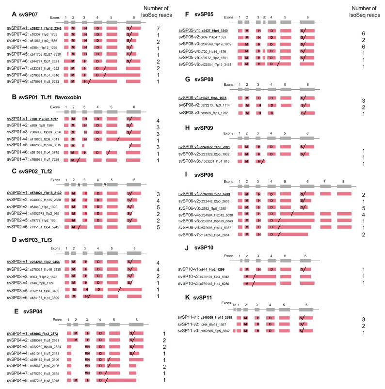
Figure 2.
Schematic diagram of structures of serine protease (SP) genes expressed in habu venom glands. svSP07 (A), svSP01-TLf1-flavoxobin (B), svSP02-TLf2 (C), svSP03-TLf3 (D), svSP04 (E), svSP05 (F), svSP08 (G), svSP09 (H), svSP06 (I), svSP10 (J), and svSP11 (K) are shown with validated transcript variants, verified as expressed in the venom gland. Initiation and stop-codons are marked with “M” and slashes, respectively. His, Asp, and Ser residues of the catalytic triad are indicated as “H”, “D”, and “S”, respectively. In svSP02-TLf2, the His residue “H” of the catalytic triad is substituted by Arg (R). Numbers of transcript variants are shown on the right. The original transcripts predicted by the gene models are underlined.

