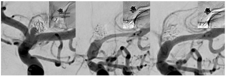
Figure 1.
Subsequent imaging findings of the symptomatic patient from uneventful implantation (left), to clinically manifest vasospasm (middle, three weeks post procedure) and after the first session of intra-arterial (i.a.) treatment (right). Note the change in caliber of the proximal and distal landing zones (black lines in each radiogram underline the change of caliber of the implanted flow-diverting stent (FDS)). The left image demonstrates normal calibers of vessel and implanted FDS approximately 30 min after the procedure. The middle image reveals high-grade tandem stenosis of the terminal internal carotid artery (ICA) and the M1 segment, caused by subacute vasospasm resulting in severe stent compression. The right image shows a significant increase in caliber immediately after i.a. spasmolysis, accompanied by clinical recovery of the patient.

