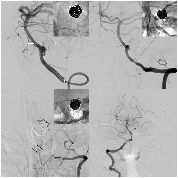
Figure 2.
Clinically inapparent case of vasospasm-associated occlusion of a left-sided, dominant vertebral artery after FDS implantation for treatment of a de novo aneurysm arising from the posterior inferior cerebellar artery (PICA) orifice. Upper row: The left image shows the left V4 segment carrying a broad-based, de novo aneurysm (a) evolving in close proximity to a previously coiled PICA aneurysm. Note the radiogram showing the optimally implanted, well-unfolded FDS, which intentionally spared the VBA confluens aiming to preserve the hypoplastic right vertebral artery as a potentially important collateral vessel (white arrow: wash-out caused by inflow from the right-sided V4). The right image shows the early follow-up DSA five weeks after implantation. Note the short, high-grade stenosis along the proximal landing zone. The radiogram detail in the upper right corner illustrates compression of the stent (black lines), additional neointimal hyperplasia (white area bordering the stent), and the residual lumen (gray area). The patient complained of recurrent episodes of cervical–occipital headaches on the left side, but otherwise remained asymptomatic.Inferior row: control angiogram and non-enhanced device image five months after treatment. The left V4 segment is occluded, and the V3 segment is reduced in size. Note the re-unfolded FDS in the radiogram (upper right corner). The formerly hemodynamically non-essential right-sided vertebral artery now independently supplies the posterior fossa. The patient did not experience a neurological deficit at any time.

