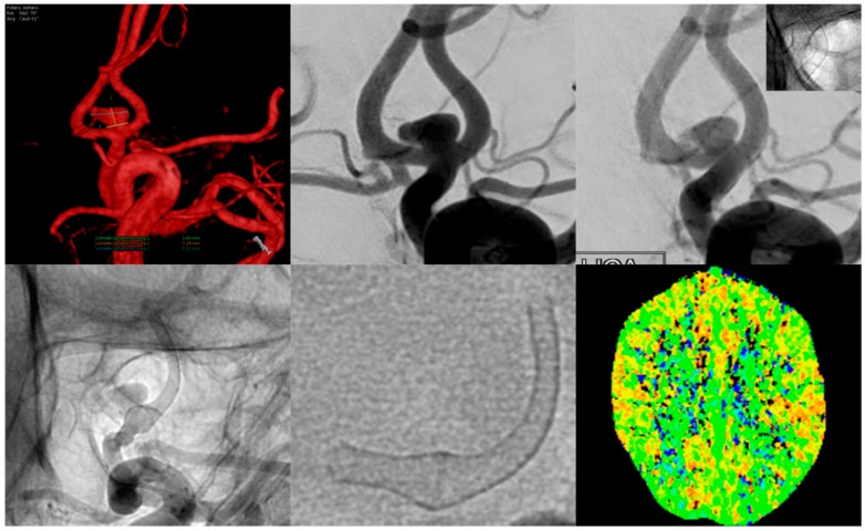
Figure 3.
Example of a patient suffering from migraine-type left-sided headache for approximately two weeks, beginning three weeks after flow diverter implantation.The upper row shows a three-dimensional (3D) rotational angiogram (left) and the conventional working projection (middle) of the AcomA-complex with the aneurysm mainly being supplied by the left A1 segment. The right image shows normal calibers of the vessel and FDS after implantation.Inferior row: Conventional, non-subtracted angiogram (left) of the left ICA shows a moderate–high-grade tandem stenosis of the proximal and distal landing zones (A1 and A2 segment of the left ACA) caused by vasospasm. The middle image shows the FDS in free projection; note the normal caliber along the aneurysm-bearing segment and the marked narrowing of the proximal and distal landing zones. CT perfusion imaging (right image) revealed no significant decrease in perfusion of the left ACA territory.

