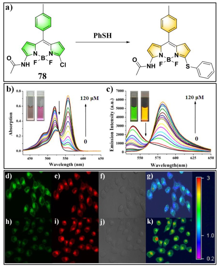
Figure 28.
(a) Structure and reaction of probe 78 for PhSH. (b) Absorption and (c) emission spectra of 78 in the presence of PhSH with different concentrations. (d–k) Confocal fluorescence images of living HeLa cells: (d–g) cells loaded with probe 78; (h–k) PhSH-incubated cells loaded with probe 78; (d,h) green channel images (500–550 nm); (e,i) red channel images (570–620 nM); (f,j) bright-field images; (g) ratio image merged from (d,e); (k) ratio image merged from (h,i). Reproduced with permission from Reference [156]; copyright 2017 Elsevier B.V., New York, NY, USA.

