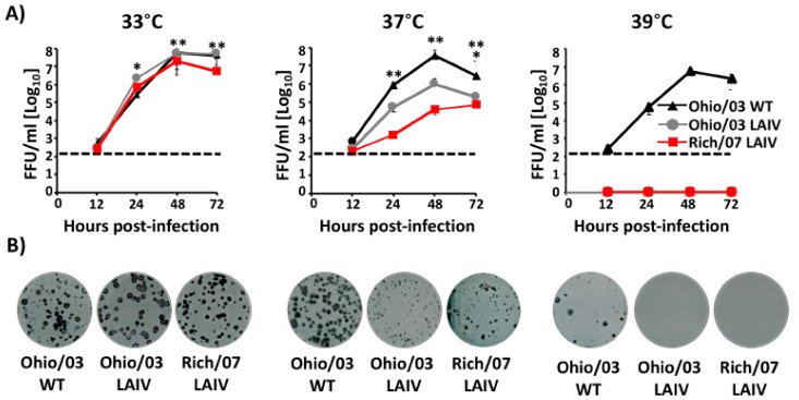
Figure 4.
In vitro characterization of Ohio/03 and Rich/07 LAIVs: (A) Multicycle growth kinetics: MDCK cells (12-well plate format, 5 × 10⁵ cells/well, triplicates) were infected (multiplicity of infection (MOI) 0.001) with Ohio/03 WT, Ohio/03 LAIV, or Rich/07 LAIV and incubated at 33 °C (left), 37 °C (middle), and 39 °C (right). Tissue culture supernatants from infected cells collected at 12, 24, 48, and 72 h p.i. were used to evaluate the presence of viruses by immunofocus assay (FFU/mL) using an anti-NP monoclonal antibody (HB-65). Data represent the means +/- SDs of the results determined in triplicate wells. Dotted black lines indicate the limit of detection of the assay (200 FFU/mL). Lines below the limit of detection represent non-detected virus. p < 0.05: * Ohio/03 WT vs. Ohio/03 LAIV; ** Ohio/03 WT vs. Rich/07 LAIV. (B) Plaque assay: MDCK cells (6-well plate format, 1 × 10⁶ cells/well) were infected with Ohio/03 WT, Ohio/03 LAIV, or Rich/07 LAIV and incubated at 33 °C (left), 37 °C (middle), and 39 °C (right). The plaque phenotype was analyzed at 72 h p.i. by immunostaining with the anti-NP monoclonal antibody HB-65.

