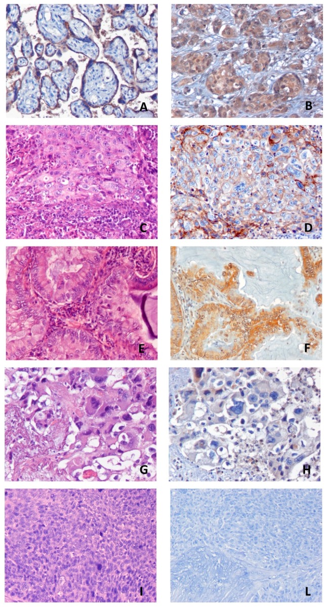
Figure 1.
Representative images of immunohistochemistry (IHC) positive controls (A,B) and tumor samples from the Nivolumab Cohort (C–L). (A): positive PD-L1 control (placenta); (B): positive B7-H4 control (breast carcinoma); (C,D): Hematoxylin and eosin (H&E) and IHC positive staining of PD-L1; (E,F): H&E and IHC positive staining of B7-H4; (G,H): H&E and IHC negative staining of PD-L1; (I,L): H&E and IHC negative staining of B7-H4. All the images are at magnification 20×.

