
Figure 1. PrEP discontinuation alters mucosal immune cell abundance and phenotype.
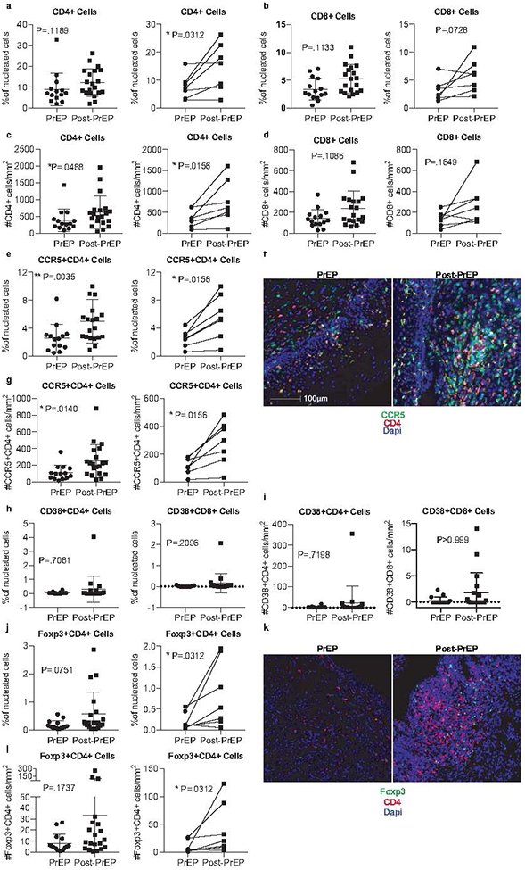
Formalin-fixed paraffin-embedded (FFPE) cervical biopsies were stained for CD4+ cells (a,c), CD8+ cells (b, d), CCR5+CD4+ cells (e, g), CD38+CD4+ or CD38+CD8+ cells (h, i), and Foxp3+CD4+ Tregs (j, l). The percentage of positive cells as frequency of total nucleated cells in whole tissue scans was determined from unmanipulated images (representative images shown in f and k). Additionally, we report the number of cells per mm2 of tissue. Results from a cross-sectional analysis or longitudinally matched pairs are shown, with lines connecting matched pairs. Cross sectional analyses are adjusted for menstrual phase, recent sex, and hormonal contraceptive use. %CD4+ cells and %CD38+CD4+ were also adjusted for BV. Thirty-five cervical biopsies were available for cross-sectional analysis: 15 women had collection at the PrEP stop visit, while 20 women had collection 2 months after PrEP; 7 of these women had paired samples at both timepoints.