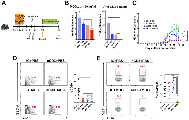
Figure 6. Anti-CD3 serves as an adjuvant to enhance oral tolerance induced by a fed antigen.
(A) Scheme for anti-CD3 (aCD3) or isotype control (IC), both given at 10 μg/mouse to enhance oral tolerance induced by MOG35-55, given at 250 μg/mouse. EAE was induced three days after the last dose of anti-CD3 or MOG35-55 and recall assay and FACS analysis performed 10 and 18 days, respectively, after immunization. (B) Ten days after MOG35-55/CFA immunization, mice were sacrificed, and spleens removed for recall assay. Splenocytes were stimulated in the presence of 100 μg/ml of MOG35-55 (specific stimulus) or 1 μg/ml of anti-CD3 (unspecific stimulus) for three days and proliferation index analyzed by H3-thymidine incorporation; n=6 mice/group. (C) EAE clinical score; n=6-7 mice/group. Only statistics for the last day of EAE follow-up (18 days post immunization) are shown. (D, E) FACS plots showing CD3+ T cell infiltration in the spinal cord (D) and Vβ11 expression on infiltrating CD3+CD4+ T cells in the spinal cord (E) 18 days after immunization; n=6-7 mice/group. Data are shown as mean ± SEM and are representative of two independent experiments. One-way ANOVA (B, D, E) or two-way ANOVA (C) were used. * p<0.05, ** p<0.01, *** p<0.001, **** p<0.0001.

