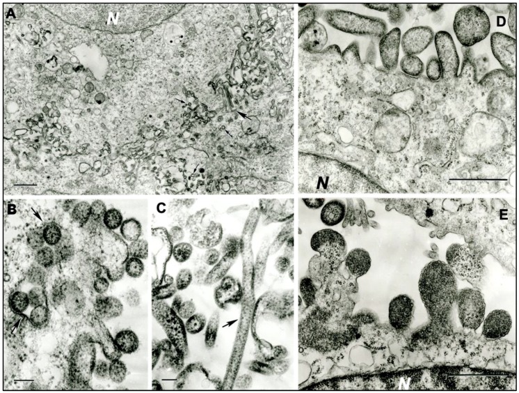
Figure 1.
Ultrastructure of MCRV (DO 159) in infected C6/36 cells, and SJCV (DO 200) in infected Vero E6 cells. (a) Multiple virus particles of MCRV forming at the cell surfaces of adjacent cells. N- cell nucleus. The large arrow shows two very long filamentous virions; small arrows show spherical virions. Bar = 1 µm. (b) Spherical virions of MCRV 130–150 nm in diameter at the cell surface. Arrows show cross-sections of the invaginations of the cell surface containing virions. Bar = 100 nm. (c) Virions of MCRV in extracellular space, a very long one (arrow) and pleomorphic. Bar = 100 nm. (d,e) Virions of SJCV forming at the surface of infected Vero E6 cells. N = fragments of cell nuclei. Bar = 1 µm.

