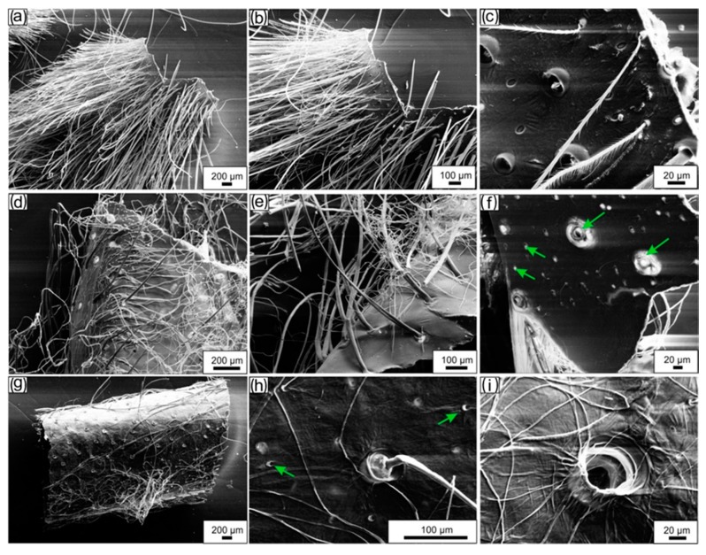
Figure 4.
SEM imagery. (a,b) A fragment of the naturally occurring molt cuticle obtained from the walking leg of C. versicolor spider represents a brush-like tube. (c) The specific structure of individual setae as well as pores on the surface of the molt are well visible on the image. (d,e) Lipid-, wax-, and pigment-free cuticle resembles the size, shape, and morphology of the molt. (f) Pores (arrows) are well visible also on the inner surface of the tube-like construct. Microwave-assisted treatment of the molt as represented in (a) leads to partial removal of the setae (g). (h,i) Pores of diverse diameter (arrows) remain visible.

