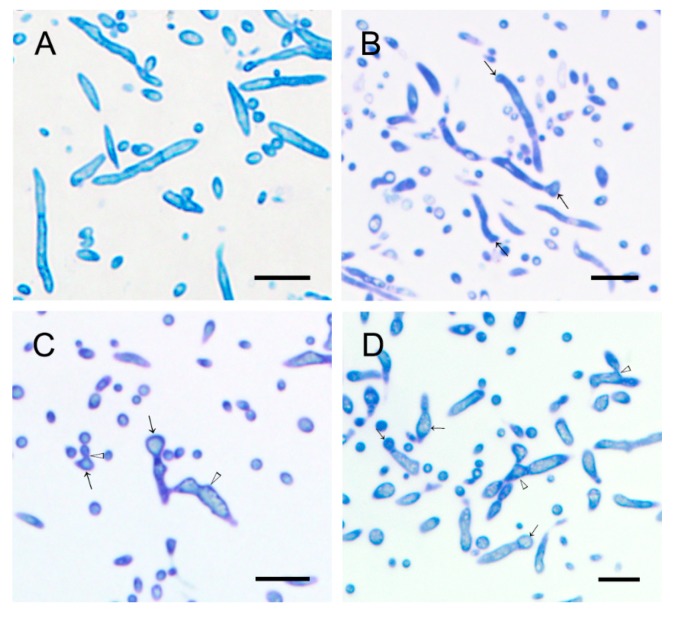
Figure 6.
Morphology of hyphae of V. mali under different concentrations of WH01 treated at 48 h. (A) At 0 mg/L treatment, healthy hyphae were exhibited. (B) At 3 mg/L treatment, morphological differences were not significant. The swollen hyphae were exhibited. At (C) 6 mg/L and (D) 12 mg/L treatment, most hyphae were malformative. Bar = 10 μm. (“→”: swollen hypha, “△” malformation of hypha).

