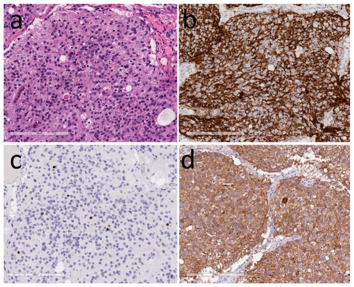
Figure 2.
Hypothalamic neurocytoma. (a) H&E identifies small round cells in abundant neuropil with a dilated neuronal axon resembling a Herring body; (b) Neurofilament highlights the neurons and neuropil; (c) TTF1 decorates some of the neurons; (d) Vasopressin is expressed by the tumor cells and highlights an axonal terminal known as a Herring body.

