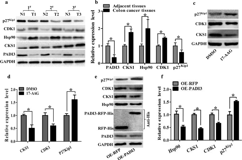
Fig. 4.
Both 17-AAG and PADI3 can suppress CKS1 expression in HCT116 cells. a Expression profile of PADI3, CKS1, Hsp90, CDK1 and p27kip1 were detected using western blot in colon cancer tissues and their corresponding adjacent tissues at the translational level, GAPDH was used to normalize the relative expression of them; b statistical analysis of western blot results in A; c 5 μM 17-AAG was used to treat HCT116 cells for 24 h, and western blot was used to measure the expression level of CKS1, CDK1 and p27kip1; d Statistic analysis of c; e Overexpression of PADI3 was performed in HCT116 cells, and western blot was used to detect the expression level of Hsp90, CKS1, CDK1 and p27kip1, overexpression of RFP as the control group; f statistical analysis of western blot results in e. GAPDH was selected as the internal control, *indicates p < 0.05 for three independent experiments analyzed by Student’s t test

