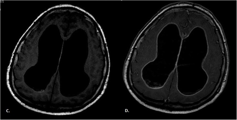
Fig. 2.

Brain MRI T1-weighted imaging without (c) and with (d) gadolinium contrast enhancement. Hydrocephalus as a consequence of disease progression. Hyper-intensity on T1 was due to the paramagnetic effect of melanin, which had stable organic free radicals inside it, resulting in shortened T1 relaxation times in typical melanotic melanoma
