Fig. 4.

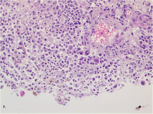
Staining of haematoxylin and eosin, enlargement 400x (f). There were many large, polymorphic cells with large nuclei and coarse chromatin. There was brown staining in the cytoplasm

Staining of haematoxylin and eosin, enlargement 400x (f). There were many large, polymorphic cells with large nuclei and coarse chromatin. There was brown staining in the cytoplasm