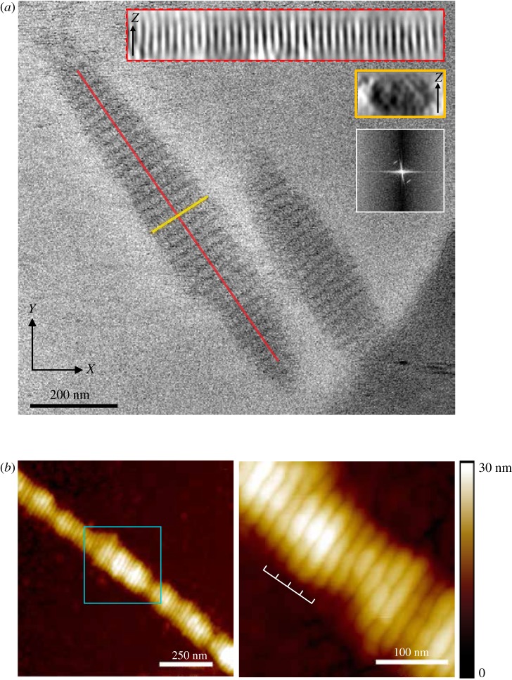
Figure 1.
Structural analysis of the SYCP3 fibre. (a) Cryo-electron micrograph of the SYCP3 fibres, showing the regular pattern of transversal striations in the fibre. The cylindrically shaped fibres measure 0.5–2 µm in length and 100–250 nm in diameter. The insets show two sections of the fibre, along and across its long axis (red and yellow boxes, respectively), and the Fourier analysis of the periodic striation of the fibre, revealing a layer line at a spatial frequency of 0.045 nm−1 (22 nm). (b) Atomic force microscopy shows that SYCP3 forms filamentous fibres that are similar to those observed by negative-stain electron microscopy [26] and by cryoEM (a). The right-hand panel shows a close-up view of the central portion of the fibre (cyan box), highlighting the presence of a transverse repeat of 22 nm.

