Figure 3.
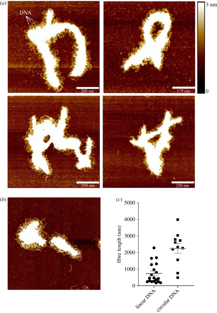
Atomic force microscopy of the SYCP3-DNA fibre. (a) Representative images of the fibrous structures formed by full-length SYCP3 in the presence of circular plasmid DNA. The DNA is visible as loops protruding from the protein core of the fibre. (b) AFM image of SYCP3 bound to linearized plasmid pUC19 DNA. (c) Scatter plot of length measurements of SYCP3-DNA fibres that incorporate either linear or circular pUC19 plasmid DNA. Individual values, mean and s.e.m. are shown.

