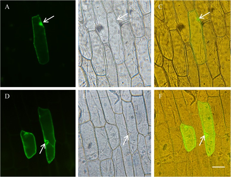
Fig. 5.
Subcellular localization of GmNHX1. Onion bulb endothelium cells expressing eGFP (a-c) and GmNHX1-eGFP (d-f) was analyzed under fluorescent microscopy, images were acquired using the 488 nM excitation (a, d) and light (b, e). Superimposed images were generated in c and f, respectively. Arrows indicate the nucleus regions. Bar = 100 μm

