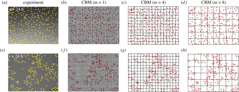
Figure 3.
Choices of compartment size in the CBM. Images of proliferation assays using cell lines 1 and 2, from figure 1c,d, are shown in (a,e), respectively. Images in (b)–(d) show various discretizations of the arrangement of cells in (a) with m = 1, 4, 8, respectively. Images in (f–h) show various discretizations of the arrangement of cells in (e) with m = 1, 4, 8, respectively. (Online version in colour.)

