
Figure 3.
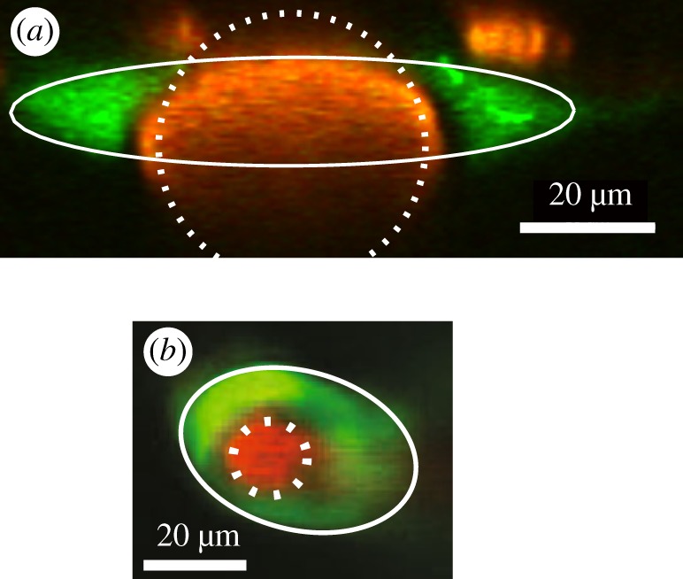
Examples of the cross-sections of PVSs (green) around arteries (red), observed in vivo. (a) Pial artery (from [21]). (b) Penetrating artery (adapted from [40]). The white lines show fits to a simple, adjustable geometric model of the cross-section of the PVS, consisting of an elliptical outer wall and a circular artery (from Tithof et al. [25]). (Online version in colour.)