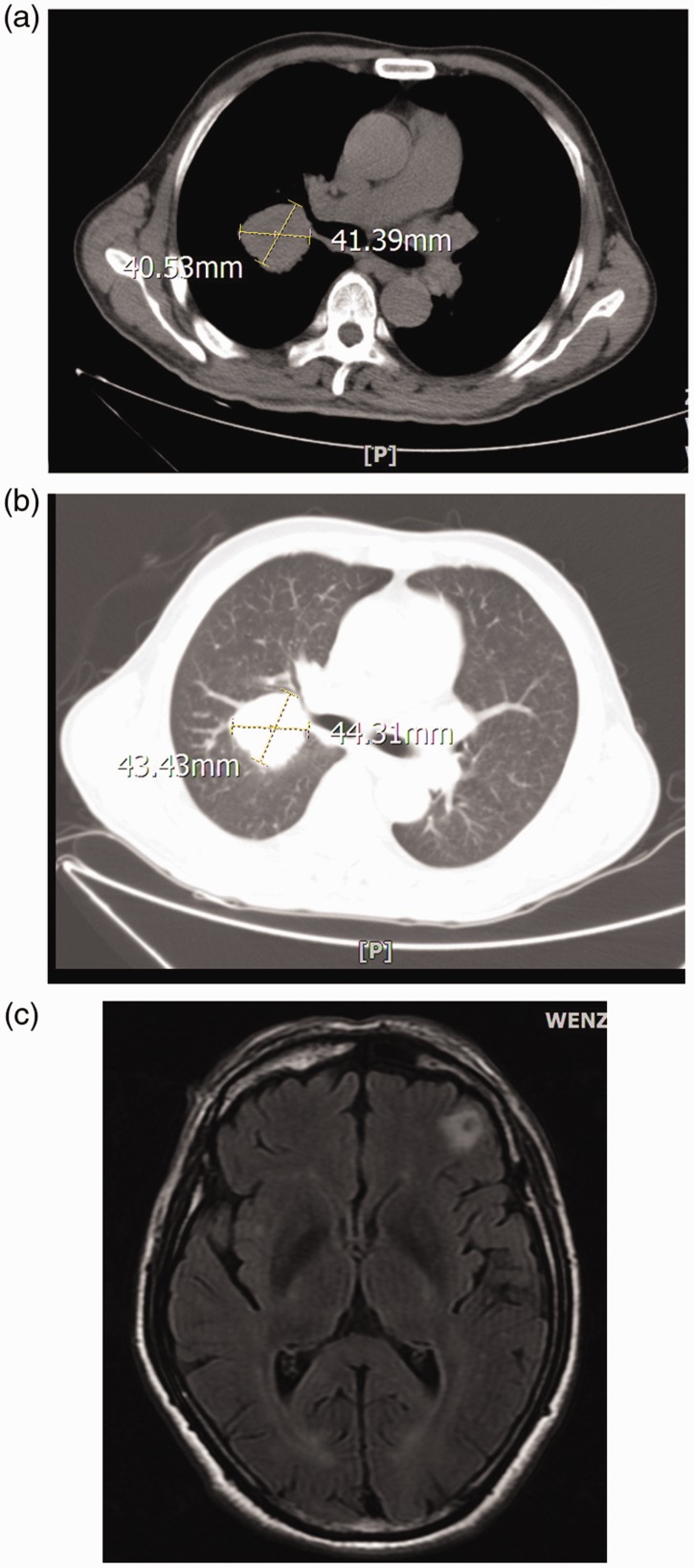
Figure 2.
Diagnostic imaging of the thorax and brain of a 62-year-old man that was admitted to the outpatient department complaining of a 3-week history of pain in the right elbow. Computed tomography scans (a and b) of the thorax showed one solid soft tissue mass (approximately 4 cm × 4 cm) with undefined margins. Magnetic resonance imaging of the brain (c) showed a round, radiolucent focus in the frontal lobe, with an irregular, opaque shadow in the central part.

