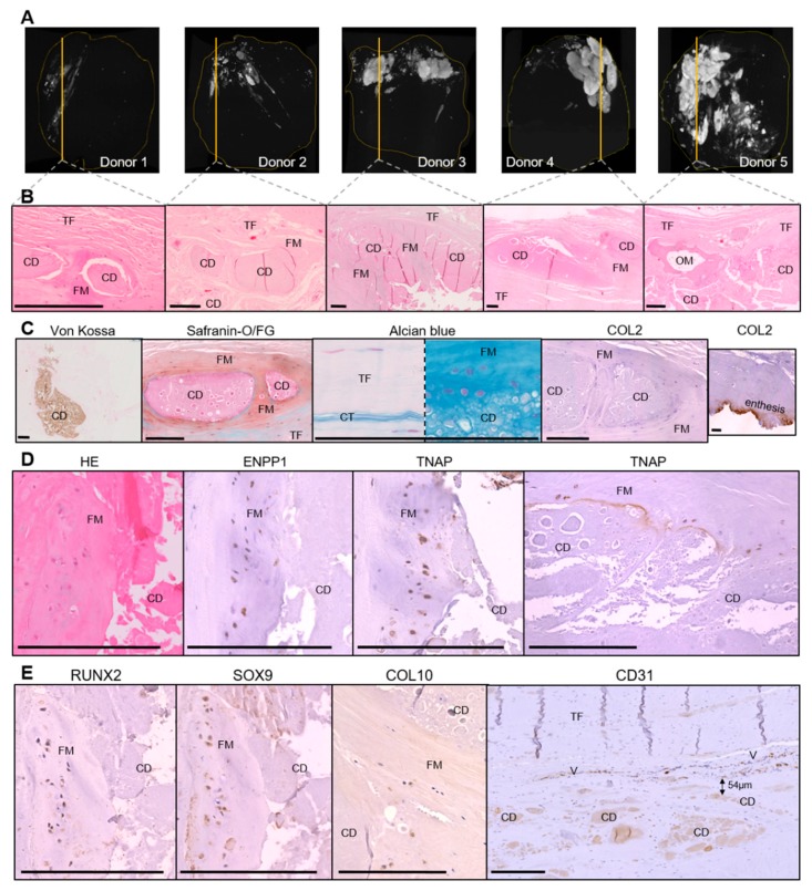
Figure 1.
Histological characterization of rotator cuff calcifications (N = 5). (A) Calcific deposits assessed by micro-computed tomography. (B) HE (hematoxylin and eosin) staining of decalcified samples (1 image for each patient). (C) Von Kossa staining on not decalcified samples and characterization of the fibrocartilaginous area with alcian blue staining, SOFG (Safranin O/Fast Green) staining, and immunohistochemical staining for COL2 (brown) with Gill hematoxylin counterstain. (D,E) Representative immunohistochemical staining for ENPP1 (ectonucleotide pyrophosphatase/phosphodiesterase 1), TNAP (tissue non-specific alkaline phosphatase), Runx2, Sox9, Col10 and CD31 (brown). TF: tendon fibers; CD: calcium deposits, FM: fibrocartilaginous metaplasia; OM: osseous metaplasia; CT: connective tissue; V: vessels. Scale bar = 200 µM.

