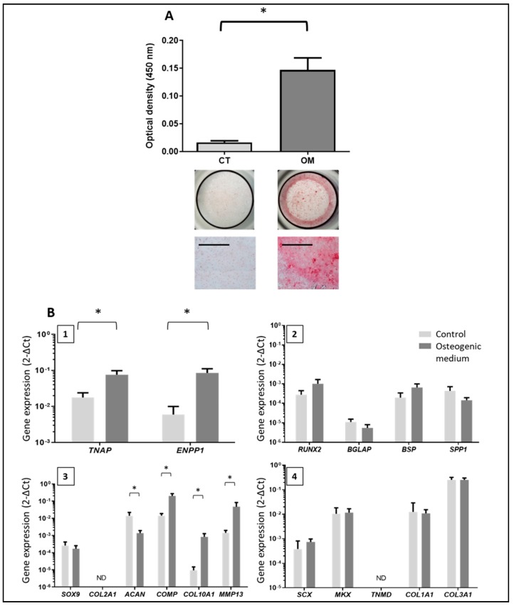
Figure 2.
Assessment of mineral deposition induced by tenocyte-like cells (TLC) after 21 days in the osteogenic medium and characterization of mineralizing cells. (A) Assessment of mineral deposition by alizarin red S staining after 21 days (N = 5). Quantification of the bound stain after solubilization with formic acid for the two conditions (reading at 450 nm) and representative images of the wells. CT: control medium; TLC: tenocyte-like cells; OM: osteogenic medium. Scale bar = 1 mm. (B) Gene expression by TLC cultured 21 days in the osteogenic medium or in the control medium (N = 3 to 5). 1: Study of TNAP (tissue non-specific alkaline phosphatase) and ENPP1 (ectonucleotide pyrophosphatase/phosphodiesterase 1) levels of expression. 2: Study of osteoblast markers: RUNX2, BGLAP (bone gamma-carboxyglutamate protein), BSP (Bone SialoProtein) and SPP1 (secreted phosphoProtein 1). 3: Study of chondrocyte markers: SOX9, COL2A1, ACAN (aggrecan), COMP (cartilage oligomeric matrix protein), COL10A1, and MMP13 (matrix metallopeptidase 13). 4: Study of tenocyte markers: SCX (scleraxis), MKX (Mohawk homeobox), TNMD (tenomodulin), COL1A1 and COL3A1. * = p < 0.05.

