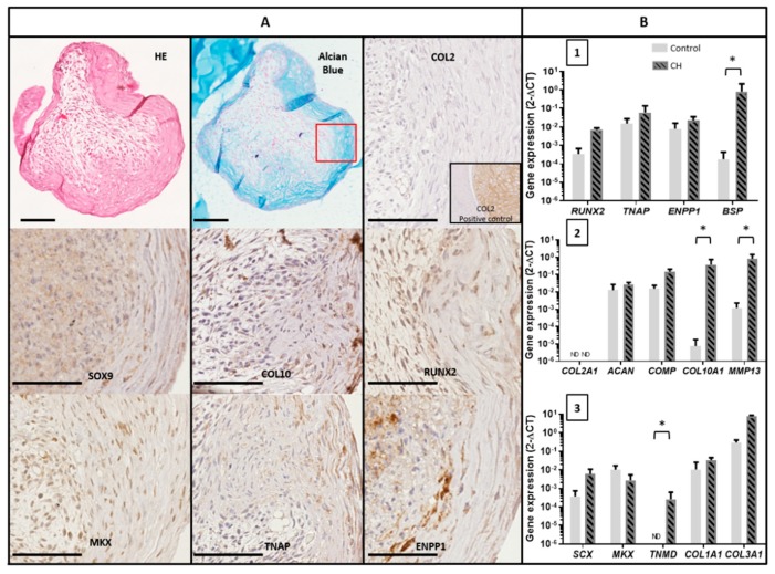
Figure 4.
Characterization of TLC cultured in chondrogenic pellets. (A) Representative images of histological and immunohistochemical staining of TLC pellets (CH medium) for COL2, SOX9, COL10, RUNX2, MKX, TNAP, and ENPP1. COL2 positive control was obtained with a chondrosarcoma sample. HE: Hematoxylin and eosin. Scale bar 100 µm. (B) Gene expression of TLC cultured four weeks in chondrogenic pellets or in cell monolayer in control medium: levels of expression of osteoblast markers (1), chondrocyte markers (2) and tenocyte markers (3) (N = 3) assessed by qPCR.* = p < 0.05.

