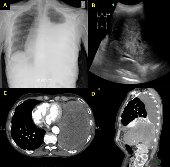
Figure 1.

(A) Posteroanterior chest radiograph reported as ‘large left-side pleural effusion causing mediastinal shift towards the right’. (B) Bedside ultrasound showing a large lobulated mass on the left side. (C) CT (axial view) showing a large lobulated soft tissue mass in the left hemithorax. (D) CT (sagittal view) of the thorax showing the mass had infiltrated through the left hemidiaphragm into the left upper quadrant.
