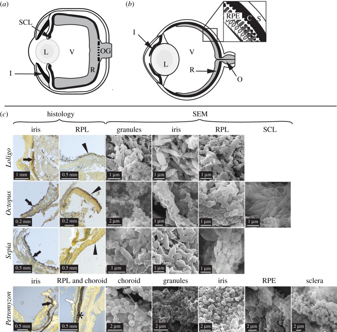
Figure 1.
Anatomy of extant cephalopod and vertebrate eyes. Schematic illustrations of (a) cephalopod eye and (b) vertebrate eye with, inset, detail of tissue layers. (c) Histological sections and scanning electron micrographs (SEM) of eyes in Loligo, Octopus, Sepia and Petromyzon. Sections are stained with Warthin–Starry; melanin appears black. All tissues show melanosome-like microbodies. C, choroid; I/black arrows, iris; L, lens; O, optic nerve; OG, optic nerve ganglia; R, retina; RPE/*, retinal pigment epithelium; RPL/arrowheads, retinal pigmented layer; S/white arrow, sclera; SCL, subciliary layer; V, vitreous humour. (Online version in colour.)

