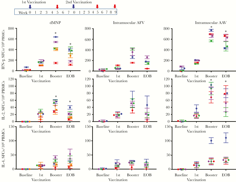
Figure 2.
Hepatitis B surface antigen (HBsAg)–specific T-cell responses after dissolvable microneedle patch (dMNP) delivery of adjuvant-free hepatitis B vaccine (AFV), intramuscular injection of AFV, and intramuscular injection aluminum-adjuvanted hepatitis B vaccine (AAV). The vaccination schedule is shown in the top panel. The booster (second vaccination) was performed at 8 weeks after the initial vaccination. Frequencies of HBsAg-specific T-helper 1 cells secreting interferon (IFN) γ (A) and interleukin 2 (IL-2) (B) or T-helper 2 cells secreting interleukin 4 (IL-4) (C) were determined with enzyme-linked immunospot assay. T-cell responses were analyzed using samples (red arrows in top panel) obtained from before vaccination (baseline), 5 weeks after the first vaccine dose (identified as 1st vaccination), 5 weeks after the second (booster) immunization, and the end of observation (EOB). Data are expressed as spot-forming cells (SFCs) per 106 peripheral blood mononuclear cells (PBMCs) and different color of dots represent levels from individual animal in each experimental group (Each color represent 5 of 7 animals receiving dMNP, 3 of 4 animals receiving intramuscular AFV and 4 of 4 receiving intramuscular AAV had anti-HBs levels ≥10 mIU/mL). There were statistically significant differences in HBsAg-specific IFN-γ and IL-2 responses between dMNP and intramuscular AAV groups at boosting (P = 0.001 and P < 0.0001, respectively) and EOB (both P < .001). *P < .001.

