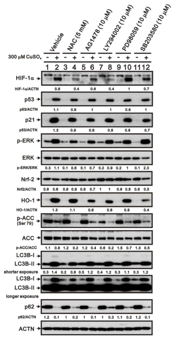
Figure 6.

Mechanism by which copper sulfate increases HIF-1α expression. HeLa cells were incubated for 8 h with the indicated compounds in the absence (−) or presence (+) of 300 μM copper sulfate. Cell lysates were then subjected to western blot analysis using antibodies against HIF-1α, p53, p21, total and p-ERK, Nrf2, HO-1, total and p-ACC, LC3B, and p62. ACTN served as the protein loading control. The results are representative of three independent experiments. Protein bands were quantified through pixel density scanning and evaluated using ImageJ, version 1.44a (http://imagej.nih.gov/ij/). The fold (shown above the bands) was normalized to the internal control (ACTN). Phosphorylated ERK and ACC after normalization of their respective total proteins are shown as fold changes.
