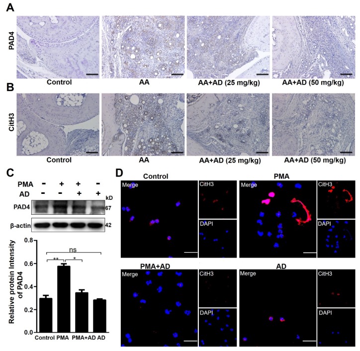
Figure 4.
AD inhibited peptidylarginine deiminase 4 (PAD4) expression and histone 3 citrullination. (A,B) Immunohistochemical analysis was performed to detect PAD4 (A) and citrullinated histone 3 (CitH3) (B) expression in the ankle joint tissue sections of each treatment group on day 37 (n = 10). Representative images are shown. (C) PAD4 expression levels were measured using Western blotting and the relative gray values were quantified using Image J. The β-actin was an internal control. Data indicate mean ± SD of three independent experiments, * p < 0.05, ** p < 0.01. (D) Phorbol 12-myristate 13-acetate (PMA)-induced neutrophil extracellular trap (NET) formation was visualized via staining neutrophils with 4’,6-diamidino-2-phenylindole (DAPI) (blue) and an anti-CitH3 antibody (red) and observed using confocal microscopy. Scale bar represents 10 μm. Representative images obtained from more than three experiments are shown.

