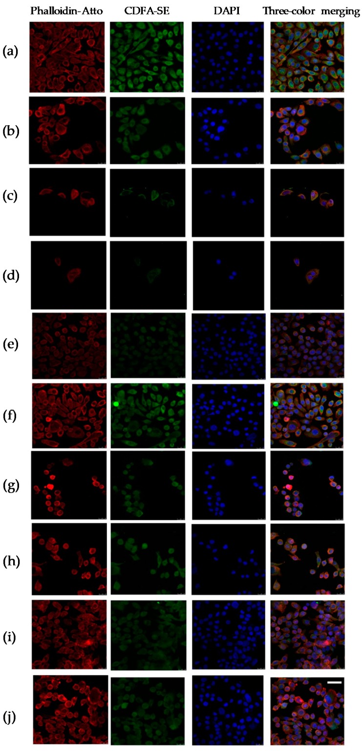
Figure 4.
Cytopathic changes in PC-3 cells induced by the control treatment (a) or the treatment with DOCE 2 nM (b); AITC 20 µM (c); AITC 20 µM + DOCE 2 nM (d); SFN 30 µM (e); SFN 30 µM + DOCE 2 nM (f); IB 30 µM (g); IB 30 µM + DOCE 2 nM (h); PEITC 4 µM (i); PEIT and C 4 µM + DOCE 2 nM (j). Confocal images show green (carboxyfluorescein diacetate succinimidyl ester, CFDA-SE), red (Phalloidin-Atto 647N), blue (4′, 6-diamidino-2-phenylindole, DAPI) and merge of three channels. The cells treated with AITC (20 µM), SFN (30 µM), IB (30 µM), PEITC (4 µM) and/or DOCE (2 nM) showed a predominantly rounded shape phenotype with DNA condensation. Scale bar = 100 µm.

