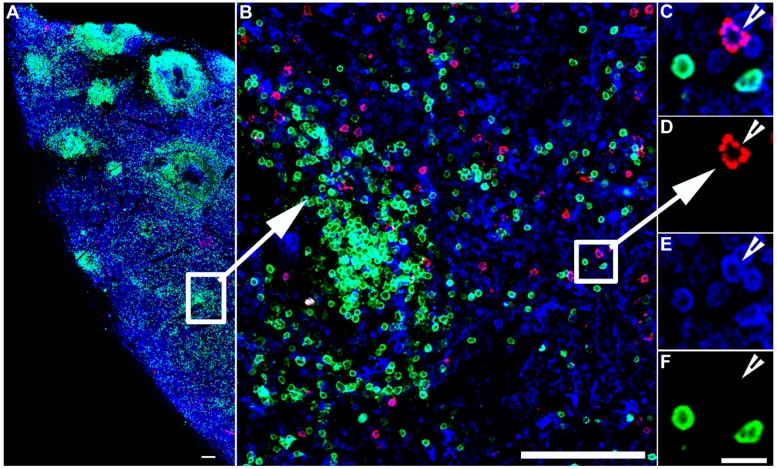
Figure 2.
IST detection of virus-specific CD8+ T cells. IST Combined with IHC in spleen sections from an SIV infected rhesus macaque. Fresh unfixed spleen section was stained with Mamu-A*01 tetramers loaded with SIV Gag/CM9 peptides detect SIV-specific CD8+ T cells (Red color), and counterstained with CD3 antibodies to label T cells blue, and CD20 antibodies to label B cells green and delineate B cell follicles. Confocal images were collected using a 20 X objective and 3 μm z-steps. (A) shows a montage of several projected confocal z-series fields. The scale bar = 100 μm. (B) shows an enlargement of the selected area in panel (A), which is a confocal Z-scan showing the distribution of tetramer+ T cells within the spleen. The scale bar = 100 μm. (C–F) are enlargements for the selected area in panel B and shows that an SIV-specific CD8+ T cell is tetramer+ (C,D), CD3+ (E), and CD20− (F), scale bars = 10 μm. Arrowheads point to a virus-specific CD8+ T cell.

