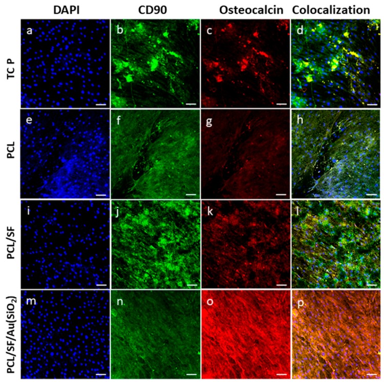
Figure 10.
Confocal microscopy images to confirm osteogenic differentiation of MSCs using MSC specific marker protein CD90 (b,f,j,n) and osteoblast-specific marker protein osteocalcin (c,g,k,o). Merged image showing the dual expression of both CD90 and osteocalcin, characteristic of MSCs which have undergone osteogenic differentiation (d,h,l,p) on TCP, PCL, PCL/SF and PCL/SF/Au(SiO2) with the nuclear staining by DAPI (blue fluorescence). Nucleus stained with DAPI (a,e,i,m) at 20× magnification (Scale bar: 50 µm).

